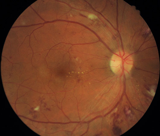
- Diabetic retinopathy.
- Retinal artery / venous occlusions. ( CRAO/BRAO & CRVO/BRVO)
- Age related macular degeneration. (DRY/WET)
- Enteral serous retinopathy.
- Hereditary/genetic retinal disorders.
- Drug toxicities like hydroxychloroquine (HCQ), ethambutol & others.
- Control of underlying systemic diseases.
- Oral and topical (eye drops) medications.
- Injections in the eye
- Retinal LASERs.
- A laser is an instrument that produces a pure, high-intensity beam of light energy. The laser light can be focused onto the retina, selectively treating the desired area while leaving the surrounding tissues untouched.
- It is used for the variety of conditions like diabetic retinopathy, retinal vein occlusions, retinal breaks or small detachments, central serous chorioretinopathy to name a few.
- There are virtually no restrictions following retinal laser, and you should be able to resume your normal activities and work schedule the following day.
- Multispot photocoagulator delivers multiple burns in a rapid predetermined sequence in the form of a pattern array produced by a scanner. The pulse duration is reduced to 10–30 ms. The aim is to optimize therapeutic effect with minimal damage to the retinal tissue. The patient is relatively more comfortable with this type of laser.
- Photodynamic Therapy (PDT) is a treatment for wet age-related macular degeneration (ARMD), a disease that involves abnormal blood vessel growth in the macula.
- PDT with Visudyne can stabilize vision and significantly reduce the risk of vision loss in certain patients with wet ARMD. Additionally, PDT with Visudyne has been proven to help slow abnormal vessel growth in patients with wet ARMD.
- Often, a diagnostic study like fluorescein angiography or OCT macula scan is performed during follow-up visits to check the retina and macula for any residual or recurring active leakage. These diagnostic tests will help the doctor to make a good, informed decision as to whether to recommend further augmentations to the PDT laser.
- Treatment of diabetic retinopathy
- Treatment of retinal tears
- Treating weak retinal areas before LASIK surgery
- Treating vascular occlusions
- Treating Central Serous Retinopathy (CSR)
- and many others
- Retinal lasers are usually done in the Out Patient Department (OPD) basis
- NO ADMISSION IS NEEDED
- Patients can walk out of the clinic almost immediately after the procedure
- A drop or 2 of an anaesthetic solution may be instilled in the eye prior to the procedure to numb (anaesthetise) the cornea after which a focusing lens is placed over the eye. the doctor the treats the affected area with green laser light and the focusing lens is removed.
- Another method of doing the LASER is by wearing a headband and using a non-contact lens to focus the laser. different methods are used for different indications
- As the laser is just light entering the eye, there is no cut/wound or opening in the eye
- The patients can immediately wash their face/eyes and there are no post-procedure restrictions
- The doctor may/may not start eye drops depending upon the indication of LASER
Intravitreal drug delivery is a method of treatment of many retinal diseases, commonly including AMD, Diabetic Retinopathy, and Retinal Vein Occlusions.
- There are several advantages of direct injections but the most important is to achieve required dose at the retina. It also increases the duration of effect of the medicine.
- The injections are absolutely painless and there is nothing to be anxious about. Majority of our patients have a smile post injection.
- The procedure is done in the operation theatre/sterile room.
- Few drops of the anaesthetic are instilled into the eye to adequately numb the surface after which the medicine is injected into the eye through the white part using a very tiny needle.
- The whole procedure takes just a few seconds.
- There is no patch after the procedure.
- Though there is no patch, patients are requested to avoid contact with dust, water for 1-2 days to reduce the chances of infection
- They can resume work the same day or at most the next morning
- One can do all routine activities like reading, writing and watching television on the same day itself
- One may also resume office the next day.
Investigations
Optical Coherence Tomography(OCT)
- The next generation 3D Optical coherence tomography system. It is a non-invasive, noncontact, trans pupillary imaging technology, with ultra-high speed 27,000 axial scans per second which allow 50 times faster data acquisition in practice and high-resolution 5-µm axial and 15-µm transverse resolution in tissue. Fourier domain OCT technology allows the systems advanced clinical protocols to be used for ocular examination with greater resolution and clarity.
- The OCT uses light waves to make a map of the retina, anterior chamber, cornea to show up any damaged areas. It uses an optical principle known as low coherence interferometry to scan across the targeted eye structure and generate an image of the same.
- The procedure requires you to sit in front of the OCT machine and place your chin on chin rest and forehead touching the head support. You are required to focus at a given target and keep your eye still while the scan is being performed.
- The layers within the retina can be differentiated and the retinal thickness can be measured. Also, the change from the previous visit, the effect of treatment can be analysed.
- The optic disc and nerve fiber layer can be assessed and guided progression analysis provides valuable information in the management of Glaucoma
- B SCAN is a two-dimensional imaging system which utilizes high-frequency sound waves ranging from 8-10 MHz. It is used for imaging of intraocular structures and giving information about the status of the lens, vitreous, retina, choroid, and sclera.
- B-scan ultrasound is most useful when direct visualization of intraocular structures is difficult or impossible in cases like - lid problems (eg, severe edema, partial or total tarsorrhaphy), keratoprosthesis, corneal opacities (eg, scars, severe edema), hyphema, hypopyon, miosis, pupillary membranes, dense cataracts, or vitreous opacities (eg, haemorrhage, inflammatory debris).
- In many instances, ultrasound is used for diagnostic purposes even though pathology is clinically visible. Such instances include differentiating iris or ciliary body lesions; ruling out ciliary body detachments; and differentiating intraocular tumours, serious versus haemorrhagic choroidal detachments, rhegmatogenous versus exudative retinal detachments, and disc drusen versus papilledema.
- A-scan mode can be used for the measurement of an axial length of the eyeball required for calculation of the lens power before cataract.
FFA (FUNDUS Fluorescein Angiography)
This investigative procedure comprises of injecting a dye -FLUORESCEIN into one of the veins in your arm and either taking rapid serial photographs of its passage within the delicate blood vessels of the eye in the retina and choroid, using a digital Fundus camera or HRA or rarely examining the inside of your eye with an INDIRECT OPHTHALMOSCOPE using appropriate filters.
Indocyanine Green Angiography (ICG)
This investigative procedure comprises of injecting a dye -INDOCYANINE GREEN into one of the veins in your arm and either taking rapid serial photographs of its passage within delicate blood vessels of the eye in the retina and choroid, using a digital Fundus camera of HRA using appropriate